Portal scintigraphy is used to diagnose portosystemic shunts in dogs and cats. The portal system consists of blood vessels that collect blood from the stomach and intestines, sending that blood through the liver as it returns to the heart. The liver is thus the first organ to receive nutrients and toxins absorbed by the intestines.

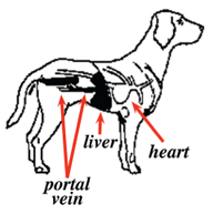
Schematic drawing of the normal portal circulation of a rectally administered material. Notice that the material is initially transported to the liver.
A portosystemic shunt is an abnormal connection between the portal circulation and the heart. This shunt is usually an abnormal blood vessel that exists from birth (congenital). Occasionally the abnormal blood vessel may develop during the animal’s life (acquired) because of an ongoing liver disease. In either case, the shunt vessel allows the blood from the gastrointestinal tract to bypass the liver and go directly to the heart and systemic circulation. As a result, the liver becomes malnourished, and the body is exposed to toxins normally removed by the liver.
The clinical signs in dogs and cats with portosystemic shunts vary highly, but they usually involve stunted growth and central nervous system signs from the presence of high levels of toxins in the blood stream. These central nervous system signs may include intermittent disorientation, pacing, circling, tremors, and even seizures. While special blood tests (e.g., serum bile acids or fasting ammonia concentrations) are useful in screening for portosystemic shunts, an imaging study is needed to confirm the diagnosis.
To perform portal scintigraphy, a small dose of the radioactive tracer is inserted into the large intestine (rectum), and the gamma camera records the route of transit to the heart. This procedure does not require sedation and only takes a few minutes to perform.

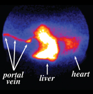
Normal study: Composite image of a normal dog showing the uptake of radioactivity into the portal vein and liver.
In a normal animal, the portal circulation absorbs the radioactive tracer and rapidly transports it to the liver. In an animal with a portosystemic shunt, the tracer bypasses the liver and travels directly to the heart. Portal scintigraphy is currently both the most accurate and safest way available to confirm the diagnosis of a portosystemic shunt. With specialized software, one can also calculate the percentage of blood that flows through the shunt vessel around the liver (the shunt fraction).
To prepare an animal for portal scintigraphy, the owner withholds food after midnight the night before and water after 6 AM the morning of the study. The owner should also withhold lactulose or similar drugs 48 hours prior to the study.

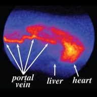
Portosytemic Shunt: Composite image showing uptake of the radioisotope from the rectum into the portal vein. Notice that the dye bypasses the liver and appears in the heart and lungs first.
